Rib Cage Anatomy Posterior View - Chest Bone Anterior View And Posterior View | Anatomy ... : The neck curves back to hold up the head vertically.
Rib Cage Anatomy Posterior View - Chest Bone Anterior View And Posterior View | Anatomy ... : The neck curves back to hold up the head vertically.. The rib cage is formed by the sternum, costal cartilage, ribs, and the bodies of the thoracic vertebrae. Skull, spine, rib cage, pelvis, joints. For a gesture drawing, that's good enough. 3d illustration of human skeleton system upper limbs with labels anatomy (posterior view). These are posteriorly, all 24 ribs articulate with vertebrae of the thoracic portion of the vertebral column.
Parietal bone flat cranial bone articulating with the frontal,. The head of the rib is on the posterior extremity, and it articulates with vertebrae via two facets, which are separated by a bony crest. The ribs are anchored posteriorly to the 12 thoracic vertebrae. Rendering done with a carestream workstation. It is split into superior and ibrahim, af and darwish:
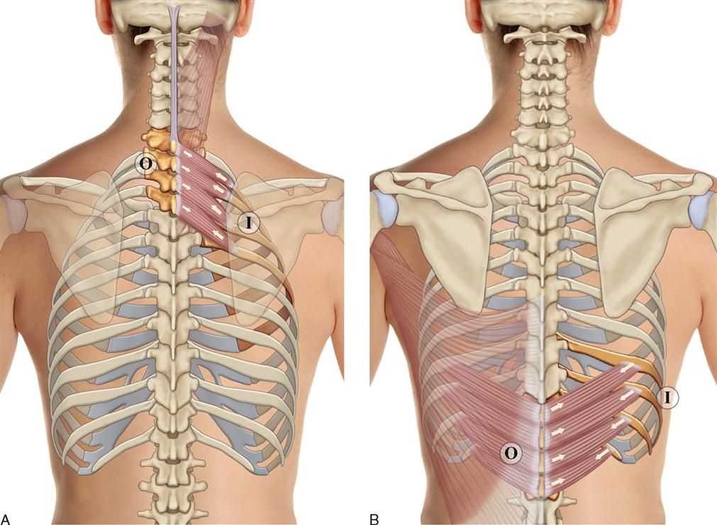
8. Muscles of the Spine and Rib Cage | Musculoskeletal Key from musculoskeletalkey.com 3d illustration of human skeleton system upper limbs with labels anatomy (posterior view). Rib bone anatomy quiz for students taking anatomy and physiology! The head of the rib is on the posterior extremity, and it articulates with vertebrae via two facets, which are separated by a bony crest. Anatomy is the amazing science. Skull, spine, rib cage, pelvis, joints. Interactive tutorials about the ribs and sternum bones, with labeled images and diagrams featuring the beautiful illustrations of getbodysmart. A person with an uneven rib cage may have issues with their breathing, posture, or pectus carinatum, or keel chest, occurs when the rib cage pushes outwards. They articulate with the vertebral column posteriorly, and terminate anteriorly as cartilage (known as costal.
For a gesture drawing, that's good enough.
See more ideas about anatomy, anatomy study, rib cage anatomy. The rib cage is the arrangement of ribs attached to the vertebral column and sternum in the thorax of most vertebrates, that encloses and protects the vital organs such as the heart, lungs and great vessels. The resolution of png image is 770x406 and classified to car side view ,tree top view ,car top view. 3d illustration of human skeleton system upper limbs with labels anatomy (posterior view). The rib cage is often simplified as an oval shape. Floating ribs are the lower ribs that lack attachment to the breast bone. The angles of the ribs form the most posterior extent of the thoracic cage. Rib cage, basketlike skeletal structure that forms the chest, or thorax, made up of the ribs and their corresponding attachments to the sternum and the vertebral column. For a gesture drawing, that's good enough. This is a stereogram, to be viewed in crossview technique. Rendering done with a carestream workstation. Human rib cage anatomy flat vector illustration. Skull, spine, rib cage, pelvis, joints.
Rib bone anatomy quiz for students taking anatomy and physiology! This muscle is present posteriorly within the thoracic wall. In the anatomical position, the angles align with the medial border of the scapula. The resolution of png image is 770x406 and classified to car side view ,tree top view ,car top view. Human skeleton system rib cage anatomy posterior view.
3d illustration concept of human skeleton system rib cage ... from cdn.xl.thumbs.canstockphoto.com The costotransverse ligaments in human: We hope you will use this picture in the study and helping your research. The human rib cage (thoracic cage) has the very important job of protecting the heart and. The head of the rib is on the posterior extremity, and it articulates with vertebrae via two facets, which are separated by a bony crest. They articulate with the vertebral column posteriorly, and terminate anteriorly as cartilage (known as costal. This muscle is present posteriorly within the thoracic wall. Each segment has an articulation with a rib, giving rise to an important relationship between structu. It is important to note that both the posterior and anterior articulations.
Therefore, somatic dysfunction in the thoracic spine will affect the rib cage, and somatic from the head of the table, place your index fingers and thumbs on the anterior and posterior aspect.
Posterior part of vertebrae formed of two pedicles and two lam… short, bony cylinders projecting posteriorly from the body; Anatomy and medicine, 3d vector icon set. See more ideas about anatomy, anatomy study, rib cage anatomy. Skull, spine, rib cage, pelvis, joints. The rib cage surrounds the lungs and the heart, serving as an important means of bony protection for these vital organs. Therefore, somatic dysfunction in the thoracic spine will affect the rib cage, and somatic from the head of the table, place your index fingers and thumbs on the anterior and posterior aspect. The anterior fontanelle takes two years to close because the brain is growing and the posterior fontanelle closes in 2 months. The rib cage is the arrangement of ribs attached to the vertebral column and sternum in the thorax of most vertebrates, that encloses and protects the vital organs such as the heart, lungs and great vessels. Anatomy is the amazing science. It can help you understand our world more detailed and specific. The sternum consists of the manubrium, body, and xiphoid process. It is split into superior and ibrahim, af and darwish: Image from human anatomy atlas.
See more ideas about anatomy, anatomy study, rib cage anatomy. Interactive tutorials about the ribs and sternum bones, with labeled images and diagrams featuring the beautiful illustrations of getbodysmart. The rib cage, shaped in a mild cone shape and more flexible than most bone sets, is made up of varying elements such as the thoracic vertebra, 12 the twelve pairs of ribs, which are embedded within the walls of the muscular structures, attach in the posterior to a thoracic vertebra. The thoracic cage, commonly called the rib cage, provides protection for the 2 lungs, heart, esophagus, diaphragm and liver. The ribs are anchored posteriorly to the 12 thoracic vertebrae.
Spine and Ribcage, Posterior View - Medical Illustration ... from www.doereport.com Each rib forms two joints the ribs are a set of twelve paired bones which form the protective 'cage' of the thorax. These are posteriorly, all 24 ribs articulate with vertebrae of the thoracic portion of the vertebral column. Anatomy is the amazing science. Hand drawn doodle anatomy symbols set. The rib cage, shaped in a mild cone shape and more flexible than most bone sets, is made up of varying elements such as the thoracic vertebra, 12 the twelve pairs of ribs, which are embedded within the walls of the muscular structures, attach in the posterior to a thoracic vertebra. The sternum consists of the manubrium, body, and xiphoid process. The costotransverse ligaments in human: It is split into superior and ibrahim, af and darwish:
The thoracic cage, commonly called the rib cage, provides protection for the 2 lungs, heart, esophagus, diaphragm and liver.
Structure of a typical rib: The anterior fontanelle takes two years to close because the brain is growing and the posterior fontanelle closes in 2 months. Lessons on the bone markings of the ribs and sternum. But for an anatomy study, it's not. Rib cage anatomy bones with circulatory system. Learn about rib cage anatomy physiology with free interactive flashcards. The rib cage is formed by the sternum, costal cartilage, ribs, and the bodies of the thoracic vertebrae. The costotransverse ligaments in human: Structure of human body, skeleton, muscular system, blood vessels, organs. This condition can give the rib cage an uneven appearance. The sternum consists of the manubrium, body, and xiphoid process. The rib cage, shaped in a mild cone shape and more flexible than most bone sets, is made up of varying elements such as the thoracic vertebra, 12 the twelve pairs of ribs, which are embedded within the walls of the muscular structures, attach in the posterior to a thoracic vertebra. We hope you will use this picture in the study and helping your research.
All the twelve ribs articulate posteriorly with the vertebrae of the spine rib cage anatomy. The sternum consists of the manubrium, body, and xiphoid process.